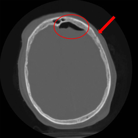

Periorbital edema and ecchymosis are frequently present. Due to the large quantum of forces involved, frontal bone fractures are often associated with other intracranial, ophthalmological and maxillofacial injuries.įS fractures can be broadly divided into four Categories: isolated anterior table fractures which comprise nearly half of the cases, isolated posterior table fractures which are relatively uncommon, combined anterior and posterior table fractures which are the most commonly seen and are severe injuries, and fractures with involvement of the NFD or NFOT.Ĭlinical presentation of FS Fractures includes the hallmark forehead lacerations, which may be accompanied by a frontal depression. Other important causes are high impact sports injuries such as martial arts, boxing injuries, interpersonal violence, extreme sports, falls from heights, and penetrating trauma from industrial accidents. This incidence has somewhat reduced with the use of seatbelts and airbags. High-velocity and high-energy impacts to the upper third of the face as seen in motor vehicular accidents are the leading cause of FS fractures. Considerable variation exists in the degree of pneumatization, ranging from extreme aeration to nonexistence of one or both of the FSs, which is seen in 5%–15% of the population. Total volume of both the FSs’ is approximately 10 cm 3. In the average adult, the FS is 30 mm tall, 25 mm wide, and 19 mm deep. Adult size is reached by 12–15 years of age and is usually characterized by two asymmetric sinuses separated by a thin bony septate plate. The FSs develop as a result of an upward extension of the anterior ethmoidal air cells which pneumatized into the frontal bone and become radiologically evident only at around 8 years of age. It is believed that this gives toddlers a harder head and protects them from fractures as they learn coordination. The NFOT is an hourglass-shaped structure, which drains secretions from the FS to the frontal recess which continues as the nasofrontal duct (NFD), which opens into the middle meatus of the nasal cavity.įSs are absent at birth and begin to form only by the age of 2 years. The sinus is lined by respiratory type of epithelium consisting of pseudostratified ciliated columnar epithelium. The floor of the sinus forms the anteromedial portion of the orbital roof and also contains the ostia or opening, through which the FS drains its mucous secretions into the nose, via the nasofrontal outflow tract (NFOT). The skull base abutting the posterior aspect of the FS is the cribriform plate of the ethmoid. The “Posterior” or “Inner” table is much thinner, just 0.1–4.8 mm in thickness, and it forms part of the anterior cranial fossa. The force needed to fracture the frontal bone is between 8 pounds, which is double the force needed to fracture the mandible and five times that needed to fracture the maxilla. This, together with the buttressing effect of its pneumatization, confers some degree of protection during trauma and allows it to resist facial fractures more than any other bone in the maxillofacial region. Its “Anterior” or “Outer” table, which forms the brow, forehead, and glabella, is strong and sturdy, with an average thickness of 4 mm, but can reach even up to 12 mm. Residual forehead contour defects and deformities.Įach FS is shaped somewhat like a pyramid, with its base directed medially, forming the intersinus septum. The immediate or acute concerns of frontal bone fractures and FS injuries revolve around:

Also of significance is the fact that as the FS is an air space containing microbial flora, that communicates with the nasal cavity, i.e., the exterior, hence FS fractures are open/compound and are prone to infections which can be potentially life-threatening. These injuries present quite a few challenges to the treating surgeon, and the ideal treatment paradigms have been debated over many years. Further, unaddressed frontal bone fractures with residual defects can leave a patient with disfiguring forehead deformities and prominent contour irregularities.įS injuries involve fractures of the frontal bone with associated involvement of the FS to varying degrees. However, because of their location and close proximity to vital structures such as the orbital and intracranial contents, these injuries can have devastating sequelae if inadequately handled or improperly managed. Fractures of the frontal bone with associated involvement of the frontal sinuses (FSs) are relatively uncommon injuries in maxillofacial trauma, comprising just around 5% of all maxillofacial injuries.


 0 kommentar(er)
